
A 2D Echo test, also known as a two-dimensional echocardiogram, is a non-invasive imaging procedure that uses ultrasound waves to create detailed images of the heart. It allows doctors to observe the heart’s structure, movement, and function in real time. This test plays a vital role in assessing how effectively the heart chambers and valves are working, and it is commonly used for both diagnosis and routine cardiac evaluation. Safe and painless, the 2D Echo provides comprehensive insights into overall heart health without the need for radiation.
A 2D Echo test is recommended when there is a need to evaluate suspected heart conditions or monitor existing cardiac issues. Common reasons include high blood pressure, history of heart disease, valve abnormalities, or unexplained symptoms such as breathlessness or fatigue. It is also advised for patients with congenital heart conditions or those who have experienced a heart attack. Additionally, individuals with risk factors like diabetes, obesity, or a family history of heart disease may be advised to undergo this test as part of preventive care.
Patients are usually referred for a 2D Echo test when they present symptoms that may indicate underlying heart problems. These include shortness of breath, chest discomfort, swelling in the legs or ankles, irregular heartbeat, dizziness, or persistent fatigue. In some cases, the test may be conducted even in the absence of symptoms, especially when routine screening or follow-up evaluation is required. Detecting subtle changes early can help prevent the progression of serious cardiac conditions.
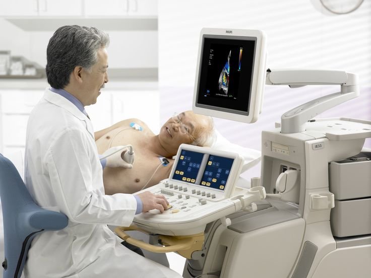
During a 2D Echo test, a handheld device called a transducer is placed on the chest to transmit ultrasound waves, which bounce off the heart structures and create moving images on a monitor. These images allow the doctor to evaluate the size and shape of the heart, the functioning of valves, and the flow of blood through the chambers. The test is usually completed within 20 to 40 minutes and does not require any special preparation. It provides accurate and detailed information that helps in identifying abnormalities such as valve disorders, heart muscle weakness, or fluid around the heart.
While the 2D Echo test itself is diagnostic, its findings are essential in guiding appropriate treatment. Based on the results, the doctor may recommend medications to manage conditions such as heart failure, hypertension, or valve disorders. In more advanced cases, interventional procedures or surgical options may be considered. The test helps ensure that treatment decisions are precise and tailored to the patient’s specific cardiac condition.
There is no recovery time needed after a 2D Echo test, and patients can resume their normal activities immediately. If any abnormalities are detected, follow-up consultations and additional investigations may be advised. Maintaining a healthy lifestyle, taking prescribed medications regularly, and attending routine checkups are crucial for effective management and long-term heart health.
A 2D Echo test is considered extremely safe, as it uses sound waves rather than radiation. There are no known significant risks or complications associated with the procedure. Some patients may feel slight pressure from the transducer during the test, but this is minimal and temporary. Overall, it is a reliable and patient-friendly diagnostic tool.
You should consider a 2D Echo test if you experience symptoms such as breathlessness, chest pain, swelling in the legs, or irregular heartbeats. It is also recommended for individuals with known heart conditions or those at higher risk due to lifestyle or genetic factors. Early evaluation through a 2D Echo enables timely diagnosis and effective management, helping to prevent serious complications and maintain optimal heart function.
WhatsApp us