Vipanchi Heart Centre
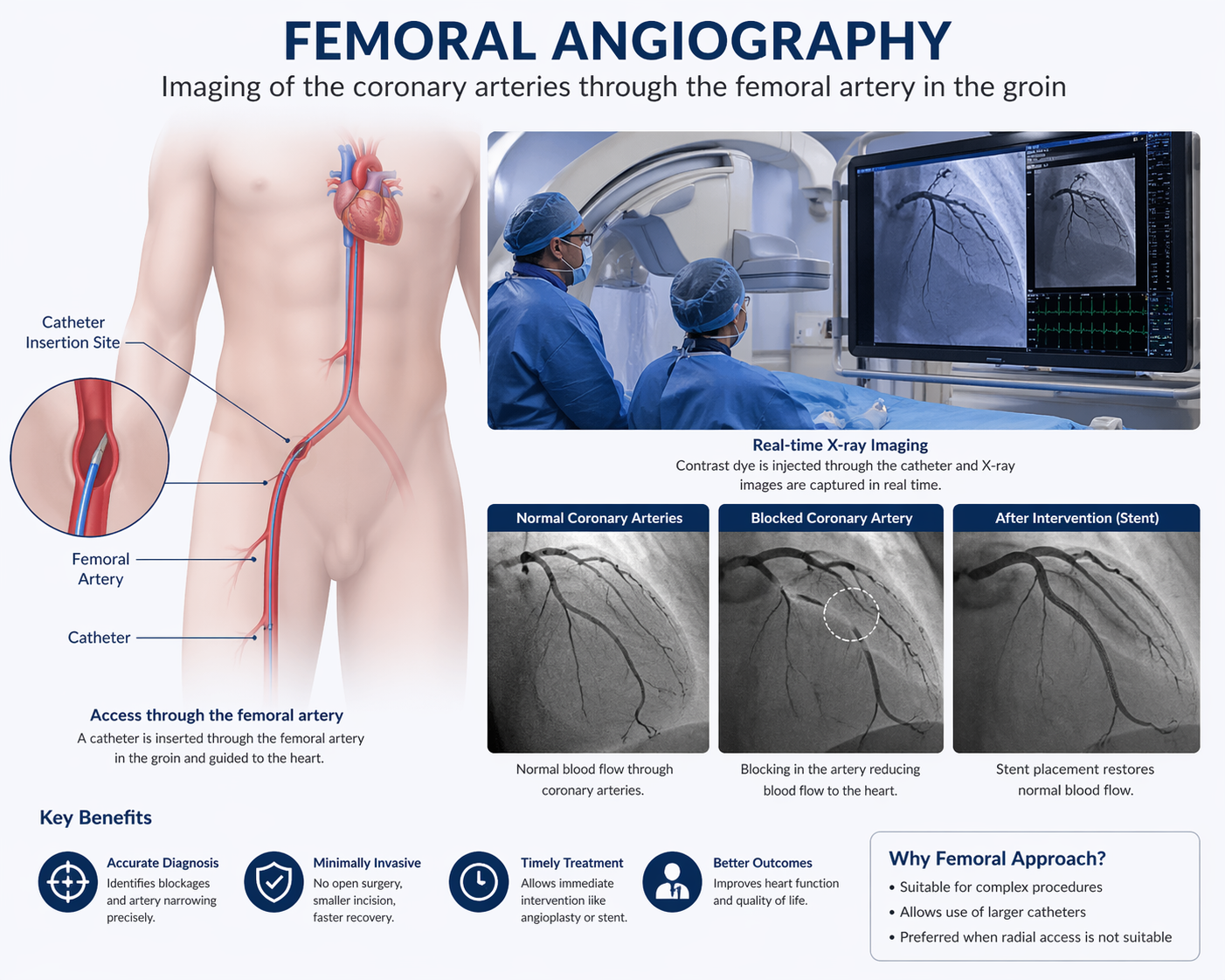
Femoral angiography is a well-established diagnostic procedure used to visualize the coronary arteries by accessing the vascular system through the femoral artery in the groin. It allows detailed imaging of blood flow within the heart and helps identify blockages, narrowing, or other abnormalities in the coronary circulation. This approach has been widely used for decades and remains an important method for evaluating complex cardiac conditions, especially when other access routes are not suitable. Femoral angiography plays a key role in diagnosing Coronary Artery Disease and guiding appropriate treatment strategies.
Femoral angiography is recommended when there is a strong suspicion of coronary artery disease or when previous tests indicate possible abnormalities. It is commonly advised for patients with persistent chest pain, abnormal ECG or stress test results, or a history of heart-related conditions. This method may also be preferred in cases where larger catheters are required or when radial access is not feasible. Risk factors such as diabetes, hypertension, smoking, and high cholesterol further increase the need for detailed evaluation.
Patients undergoing femoral angiography often present with symptoms that suggest reduced blood flow to the heart. These include chest pain or pressure, shortness of breath, fatigue, dizziness, or palpitations. In some situations, the procedure may be performed even in the absence of symptoms, particularly when routine investigations reveal potential cardiac concerns that require further evaluation.
Coronary angiography itself is a diagnostic tool used after initial assessments such as electrocardiograms (ECG), stress tests, or echocardiograms suggest possible heart disease. During the procedure, a thin catheter is inserted through a blood vessel—usually in the wrist or groin—and guided to the heart. A contrast dye is injected, allowing real-time X-ray images to capture the flow of blood through the coronary arteries. This enables precise identification of blockages, their severity, and location, which is essential for planning treatment.
Although femoral angiography is primarily a diagnostic procedure, it can be combined with therapeutic interventions if needed. If significant blockages are identified, procedures such as balloon angioplasty or stent placement can be performed during the same session. This integrated approach allows for immediate treatment and reduces the need for additional procedures.
After femoral angiography, patients are usually required to lie flat for a few hours to prevent bleeding from the insertion site. Recovery may take slightly longer compared to radial access, but most patients can return to normal activities within a day or two. Proper care of the insertion site, adherence to medications, and follow-up visits are important for a smooth recovery and long-term heart health.
Femoral angiography is generally safe, but it carries some risks, including bleeding, bruising, or infection at the groin site. There may also be a small risk of blood vessel damage, allergic reaction to the contrast dye, or irregular heart rhythms. Serious complications are rare, and the procedure is performed under expert supervision to ensure patient safety.
You should consult a doctor if you experience symptoms such as persistent chest pain, breathlessness, or abnormal findings in previous heart tests. Individuals with multiple risk factors for heart disease should also consider timely evaluation. Femoral angiography provides detailed insights into coronary health and helps in planning effective treatment to prevent serious cardiac events.

WhatsApp us